Research
Elucidating interactions between behavior-generating circuits using functional and anatomical connectomics
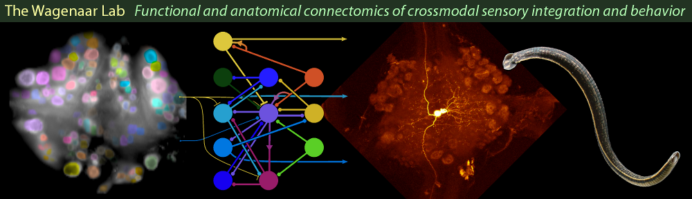
How brain activity can lead to complex and flexible behavioral outputs has fascinated neuroscientists and philosophers alike. There is mounting evidence that complex behaviors result from the activity of a multitude of simpler (sometimes competing) circuits. Yet, our understanding of even the simplest circuits remains spotty, in part because available technology has limited researchers to studying only one or a few aspects of a circuit at a time. We stand at the cusp of a revolution in recording and imaging technology that will ultimately allow us to investigate comprehensively how the fundamental biological building blocks of the human brain are constructed and fit together. Even now, the limitations mentioned no longer apply to certain less complex, more experimentally approachable brains. These provide attractive stepping stones for understanding our own complex brain. We use the relatively simple nervous system of the European medicinal leech to develop insights about how the activity of all the cells in a nervous system together produce individual behaviors from overlapping functional networks, a phenomenon that—at a much larger scale and undoubtedly with many complexities added—is also crucial to human brain function.
We perform three types of experiments:
- Recording the activity of all the neurons in a ganglion—the unit of activity in this animal’s brain—using high-resolution voltage-sensitive dye imaging as the animal performs four different behaviors: swimming, crawling, local bending, and shortening;
- Using electron microscopy to reconstruct the full connectivity pattern—the “connectome”—of the same ganglion that was imaged;
- Using electrophysiology to add functional significance to the anatomical connectome.
We have already obtained a simultaneous activity record of all the individual neurons in several ganglia as they generate several behaviors. (See our recent eLife paper.) One of these ganglia is presently being sliced and imaged in a serial blockface electron microscope (SBEM). Combining the activity record with the reconstructed connectome based on the SBEM images will establish a data set with unprecedented potential for advancing our understanding of the link between neuronal connectivity and behavior. In analyzing this data set, a particular focus will be on neurons and synaptic connections that span multiple behavioral circuits, to determine their roles in selecting behaviors.
Multisensory integration
Obtaining information from the environment to guide behavior is one of the most fundamental functions of nervous systems. Most animals combine cues from multiple sensory modalities to gain information about their environments. When individual cues are not 100% reliable, combining cues greatly aids decision making and it makes behavior more robust under variable circumstances. My lab studies the neuronal basis for such ``multisensory integration'' in the medicinal leech Hirudo verbana. Hirudo is an obligate sanguivore that preferentially feeds on mammalian blood. It lives on the bottom of shallow ponds. When a mammal steps into the water and splashes around, the leech will swim toward the source of the disturbance to find its prey. Leeches can find their prey in total darkness, relying on water-wave sensors on their skin. Remarkably, they can also find their prey using sensory stimuli alone: when placed in a shallow tank, they will swim toward a disturbance in a second tank placed above theirs but mechanically isolated from it. Under more general circumstances, cues from both modalities are available, and leeches must either combine the two modalities, or decide which one is more reliable and selectively ignore the other. The goal of my lab is to find out how their nervous system solves this challenge and produces a coherent decision for subsequent motion.
Hirudo is an ideal animal for such investigations, because of its reliable responses to visual and non-visual stimuli, the simplicity and accessibility of its nervous system, and the robustness of its responses even after extreme surgery. Importantly, its visual system is quite primitive (see below) so its mechanism for multisensory integration is likely to be close to the most basic one possible; a good starting point for discovering fundamental properties that such a mechanism must have in order to be functional at all. Thus, elucidating this mechanism in great detail will suggest general properties for mechanisms of multisensory integration in other species and motivate experiments to further explore the far more complex integration that occurs in higher brains.
Visual processing in the medicinal leech
A crucial step toward the overall goal of the lab is to improve our understanding of the neural circuits involved in visual processing. The entrypoint of the visual system of the leech consists of five pairs of primitive eyes located on the head, and seven pairs of photosensitive ``sensilla'' located around the body circumference at each of its 21 midbody segments. Neither eyes nor sensilla have image forming optics. The projections of the eyes and sensilla are known, and several specific cells in the central nervous system have been identified as receiving visual input, but a systematic exploration of the visual pathways either in the headbrain or in the segmental ganglia has not been undertaken. Yet, such an endeavor is eminently feasible in the leech, because there are only about 400 cells in each ganglion, and their anatomy and functions are strongly stereotyped. One very attractive question is whether and how the leech utilizes its 14x21 sensillar array to form a basic image of the visual world.
Techniques
The major challenge for studying how information flows through the nervous system is recording from a large number of cells at once. We will meet this challenge using voltage-sensitive dye imaging, multielectrode array recording, and serial blockface electron microscopy. The combination of these techniques will make it possible to record neural activity with single-cellular spatial resolution and single-action-potential temporal resolution and link this activity to anatomical connectivity at the single-synapse spatial scale.